Cellular Potts Model Simulator
A CPM simulator for modeling collective migration of Dorsal Forerunner Cells during zebrafish development. Cells as deformable bodies on a lattice with Gaussian filopodia, durotaxis, and contact interactions at 10-50 FPS.

Business Context
Understanding how cells collectively migrate during embryonic development is a fundamental question in developmental biology. During zebrafish embryogenesis, Dorsal Forerunner Cells migrate collectively to form the Kupffer's vesicle — the organ that establishes left-right body asymmetry. Agent-based models that treat cells as points or rigid circles miss the essential physics: cells deform, they stick to each other with type-dependent forces, and they extend probing filopodia that pull them in specific directions.
Strategic Value
The Cellular Potts Model represents each cell as a deformable body on a lattice, evolving through Metropolis Monte Carlo sampling of a Hamiltonian that balances area constraints, perimeter constraints, adhesion energy, and Gaussian filopodia motility. The filopodia model R(θ) = max{R₀ + Aⱼ·exp(-(θ-θ₀ⱼ)²/(2Wⱼ²))} creates directionally biased protrusion driving cell crawling. Real-time 2D Canvas visualization at 10-50 FPS with two-pass collision resolution. Developed at SCIAN-Lab and BNI, Universidad de Chile, supporting published developmental biology research.
The Challenge
Understanding how cells collectively migrate during embryonic development requires models that capture deformable cell shapes, contact interactions, and directed motility — not just point-particle dynamics.
Our Approach
Hamiltonian minimization balancing area constraints, perimeter constraints, adhesion energy, and Gaussian filopodia motility. Two-pass collision resolution, per-cell color coding, tissue boundary dynamics, cell proliferation.
Key Performance Indicators
| KPI | Baseline | Result | Impact |
|---|---|---|---|
| Cell Model | Point particles | Deformable bodies on lattice | Realistic shape dynamics |
| Visualization | Post-processing only | Real-time 2D Canvas at 10-50 FPS | Interactive parameter exploration |
Architecture
cpm simulator
The Biology
During zebrafish embryonic development, Dorsal Forerunner Cells (DFCs) migrate collectively to form the Kupffer’s vesicle — the organ that establishes left-right body asymmetry. Understanding this process requires a model that captures what actually matters: cells aren’t points or rigid circles. They deform. They stick to each other. They extend probing filopodia that pull them in specific directions.
The Model
The Cellular Potts Model represents each cell as a deformable body occupying multiple lattice sites. The system evolves through Metropolis-style Monte Carlo sampling — proposing site-copy events and accepting or rejecting them based on a Hamiltonian energy function that balances four competing forces:
Area and perimeter constraints keep cells from growing or shrinking unrealistically. Adhesion energy governs how strongly different cell types stick to each other. And the Gaussian filopodia model drives migration:
R(θ) = max_j { R₀ + Aⱼ · exp(-(θ - θ₀ⱼ)² / (2·Wⱼ²)) }
Each filopodium creates a directional protrusion bias — amplitude Aⱼ, preferred direction θ₀ⱼ, angular width Wⱼ. The maximum over all filopodia determines the cell’s effective reach, creating the crawling behavior observed in real DFC migration.
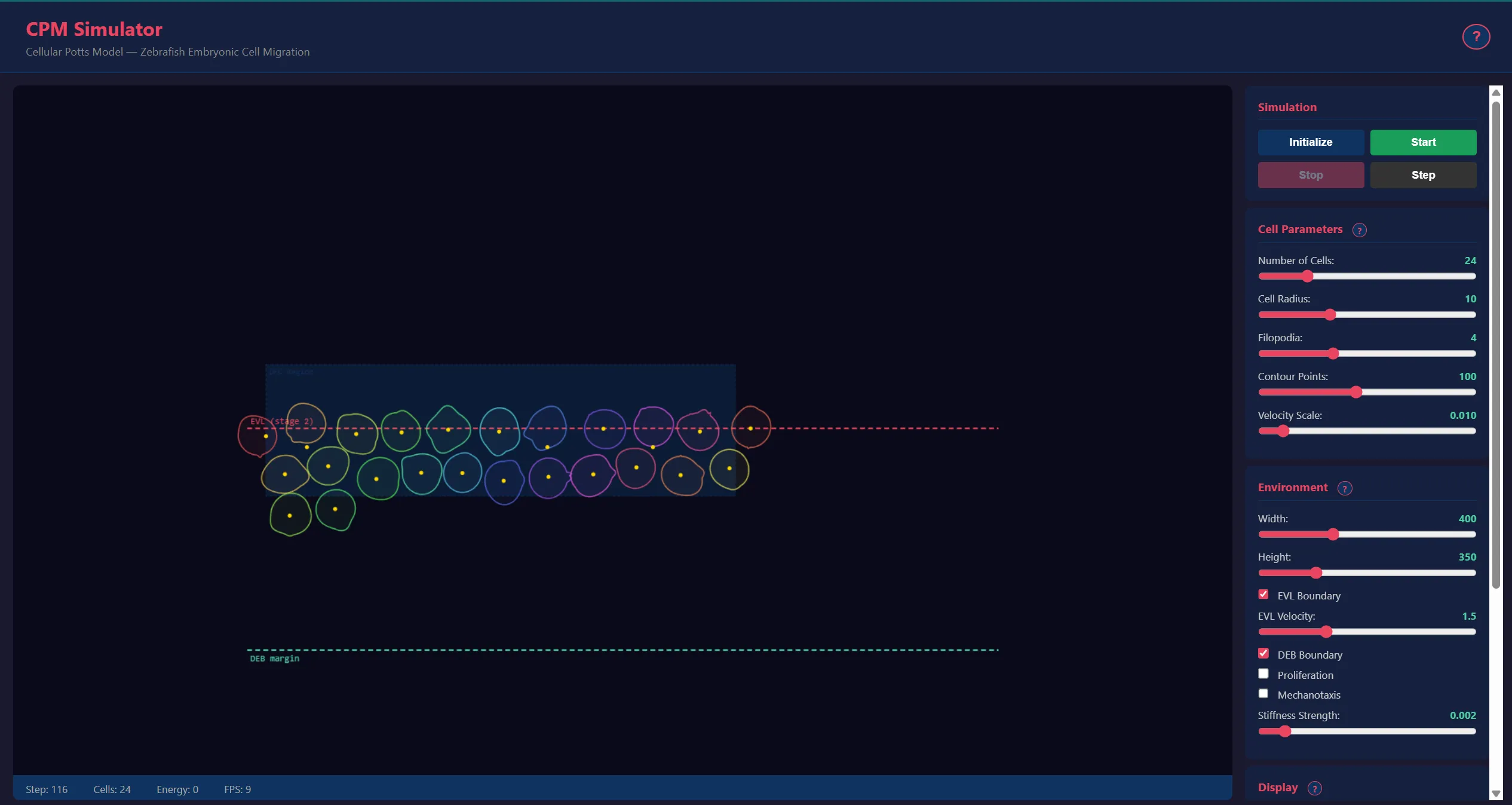
The simulation runs at 10–50 FPS with real-time 2D Canvas visualization, two-pass collision resolution, and tissue boundary dynamics (EVL and DEB layers). Developed at SCIAN-Lab and BNI, Universidad de Chile, supporting developmental biology research.
Technology Stack
Application Screenshots
Technical Diagrams

cpm biological context

cpm cell model

cpm collision passes

cpm durotaxis